

Accurate evaluation of stent encrustation patterns, such as volume distribution, from different patient groups are valuable for clinical management and the development of better stents.
Shaokai Zheng, Pedro Amado, Bernhard Kiss, Fabian Stangl, Andreas Haeberlin, Daniel Sidler, Dominik Obrist, Fiona Burkhard and Francesco Clavica.
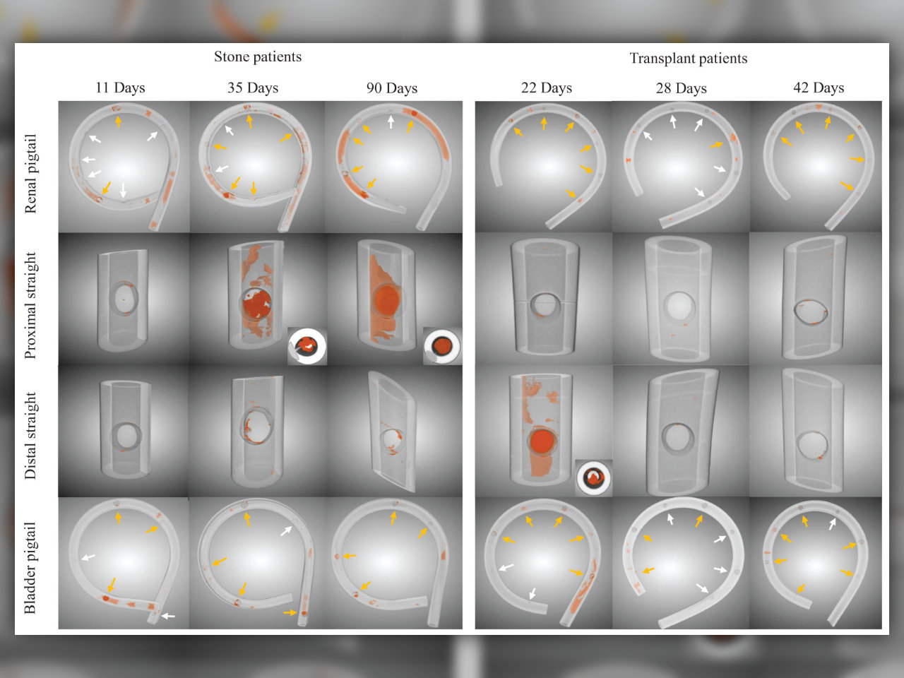
Abstract: Accurate evaluation of stent encrustation patterns, such as volume distribution, from different patient groups are valuable for clinical management and the development of better stents. This study quantitatively compares stent encrustation patterns from stone and kidney transplant patients. Twenty-seven double-J ureteral stents were collected from patients with stone disease or who underwent kidney transplantation. Encrustations on stent samples were quantified by means of micro−Computed Tomography and semantic segmentation using a Convolutional Neural Network model. Luminal encrustation volume per stent unit was derived to represent encrustation level, which did not differ between patient groups in the first six weeks. However, stone patients showed higher encrustation levels over prolonged indwelling times (p = 0.02). Along the stent shaft body, the stone group showed higher encrustation levels near the ureteropelvic junction compared to the ureterovesical junction (p = 0.013), whereas the transplant group showed no such difference. Possible explanations were discussed regarding vesicoureteral reflux. In both patient groups, stent pigtails were more susceptible to encrustations, and no difference between renal and bladder pigtail was identified. The segmentation method presented in this study is also applicable to other image analysis tasks in urology.
Keywords: Double J, ureteral stent, encrustation, stone, renal transplantation, micro CT, segmentation, deep learning.
COST is supported by the EU
Framework Programme Horizon 2020.
COST Association
Avenue Lousie 149 | 1050 Brussels, Belgium
info@enius.org
Tel: +32 (0)2 533 3800 | Fax: Fax: +32 (0)2 533 3890
Email usEuropean Network of Multidisciplinary Research to Improve the Urinary Stents (ENIUS)